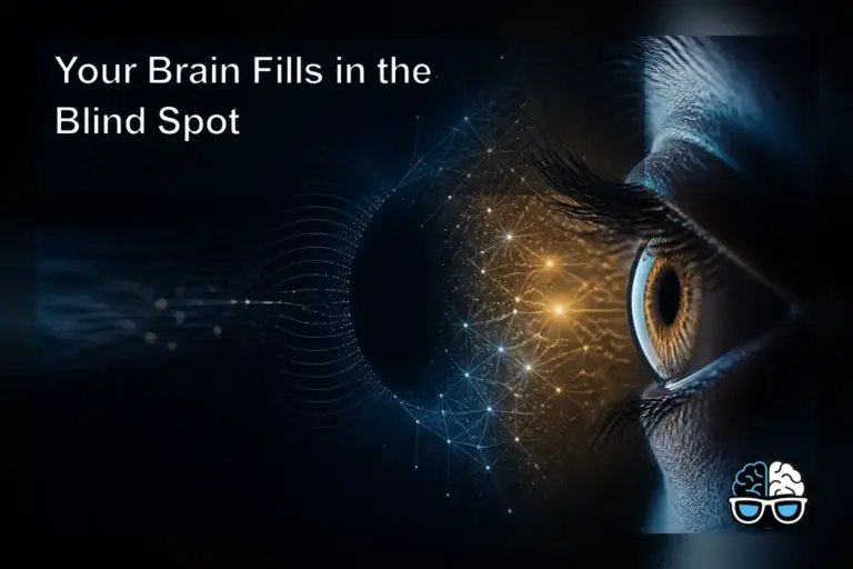
Your Brain Fills in the Blind Spot
Explains the eye's blind spot, how the brain fills in missing visual input, its location, discovery by Mariotte, and why it matters for perception.
Overview
The blind spot is also called a scotoma.
It is an obscuration of the visual field where no light is detected.
It corresponds to the optic disc, the point where the optic nerve exits the eye.
The optic nerve passes through the retina at this location.
Because photoreceptor cells are absent at the optic disc, the corresponding part of the visual field would be invisible.
The brain compensates by filling in the missing information.
It uses surrounding visual detail and input from the opposite eye.
This interpolation makes the gap normally imperceptible to conscious vision.
In medical literature the blind spot is also referred to as the physiological blind spot, the blind point, or the punctum caecum.
The resulting perceptual completion allows observers to experience a continuous visual field despite the anatomical gap.
This neural interpolation prevents the blind spot from creating a noticeable hole in what we see.
Background
The phenomenon was first documented in the 1660s by the French scientist Edme Mariotte.
At that time, the prevailing belief held that the point where the optic nerve entered the retina should be the most sensitive area of vision.
Mariotte’s experiments revealed the opposite: no light perception at that specific point.
He presented his findings to the court of King Louis XIV of France.
Later he shared the results with the Royal Society of London.
This sparked interest among natural philosophers.
His work challenged long‑standing assumptions about retinal uniformity.
It paved the way for later studies on visual processing.
The article includes a dedicated History section that outlines Mariotte’s experiments and their reception by the French court and the Royal Society.
Mariotte’s findings challenged the long-held view that the optic nerve entry point was retinally most sensitive.
His work laid the foundation for subsequent investigations into how the visual system handles incomplete information.
Key details
In humans, the blind spot lies about 12 to 15 degrees temporal to the fovea.
It is also roughly 1.5 degrees below the horizontal meridian.
Its approximate dimensions are 7.5 degrees in height.
And 5.5 degrees in width.
All vertebrate eyes possess this anatomical blind spot.
This is because the optic nerve must pass through the retina.
In contrast, cephalopod eyes evolved independently.
They route the optic nerve behind the photoreceptor layer.
This leaves no interruption in the retinal surface.
Under dark‑adapted conditions, the rod‑dominated peripheral vision can produce a central night blind spot.
The foveal cone‑rich area remains less sensitive to low light.
The blind spot can extend over the entire retina as an angioscotoma.
The entry contains a section titled “Blind spot test” that describes methods for detecting the scotoma.
It also features a “Night blind spot” section explaining how rod‑dominated peripheral vision can affect low‑light perception.
The “See also” list includes related concepts such as Bias blind spot, Filling‑in, Horizontal eccentricity, Acute idiopathic blind spot enlargement syndrome, and Angioscotoma.
Finally, the article concludes with a References section that cites the sources used for the information presented.
The presence of the blind spot in all vertebrates points to a shared anatomical constraint in vertebrate eye design.
In cephalopods, the optic nerve approaches the photoreceptors from behind, preventing a retinal break.
The angular measurements are expressed in degrees of visual angle, reflecting the size of the gap on the retina.
Why it matters
Understanding the blind spot shows that perception is an active construction by the brain.
It is not a direct copy of retinal input.
The filling‑in process relies on neural interpolation.
It uses contextual information from surrounding areas.
It also uses input from the opposite eye.
This mechanism explains why individuals are generally unaware of the gap despite its anatomical absence.
Demonstrating the blind spot provides a clear example of the brain’s compensatory strategies for missing visual data.
It also relates to broader principles of sensory processing.
It shows how neural circuits fill in incomplete information.
Recognizing how the brain fills in the blind spot highlights the constructive nature of vision.
It also emphasizes the role of neural interpolation in everyday sight.
The brain’s filling‑in activity exemplifies how perceptual systems use prior knowledge to fill missing sensory data.
This process is consistent with theories of predictive coding in visual perception.
Knowledge of the blind spot aids in diagnosing certain visual field defects and understanding normal vision.
External References
For more detailed information, consult the Wikipedia entry on the blind spot: https://en.wikipedia.org/wiki/Blind_spot_(vision)
The primary source for this article is the Wikipedia entry on blind spot (vision) available at the provided link.
Readers can follow the link to explore the full article and its references.
Sources